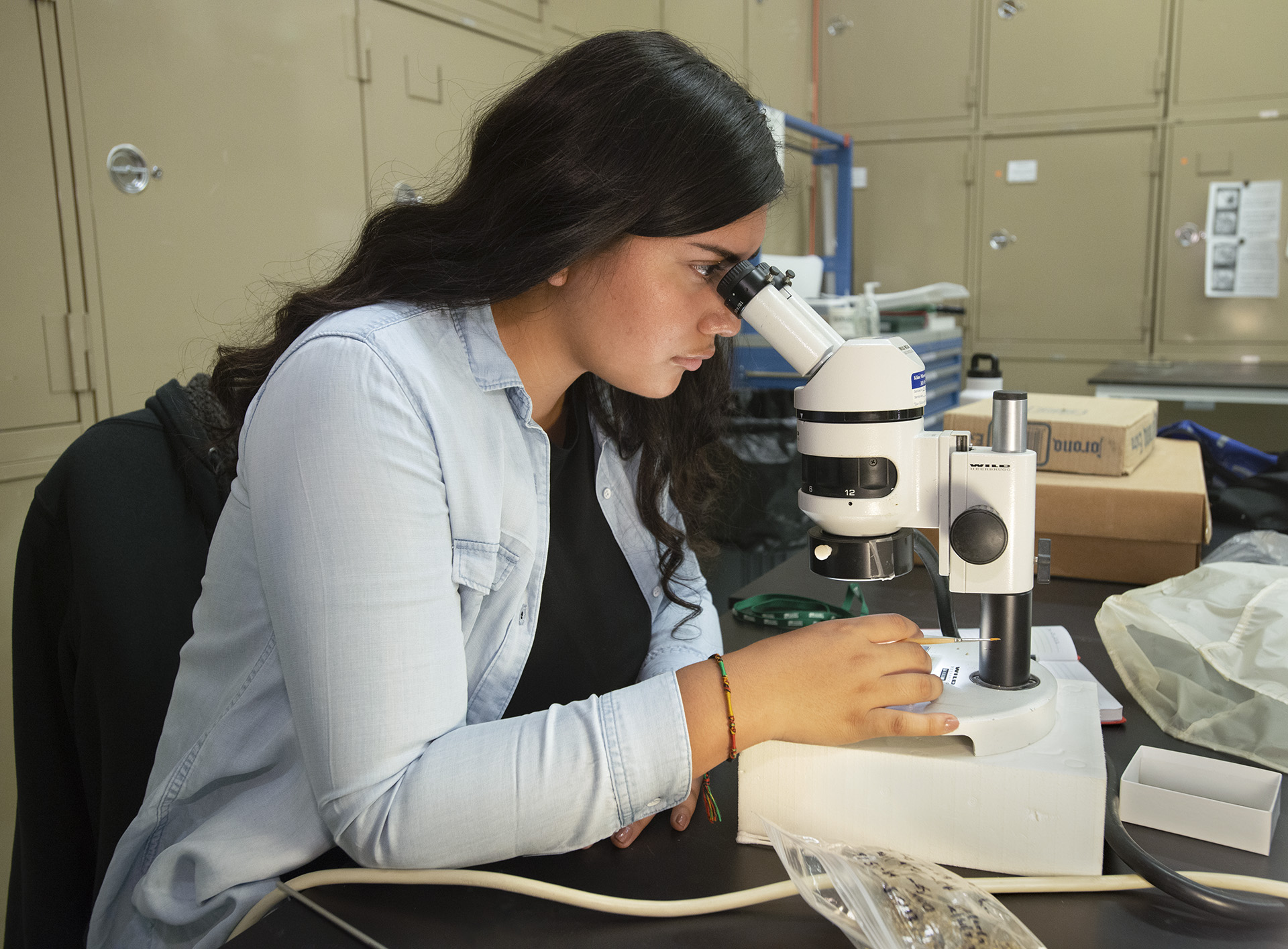
The Digital Research Lab is a research facility that uses cutting-edge visualization methods to reveal the anatomical structure of Museum specimens
The visualization methods employed in the Digital Research Lab include imaging, modeling, virtual reconstruction, and animation. The lab is focused on helping our curators address questions related to the anatomical structure of organisms, both fossil and modern. The noninvasive and nondestructive digital methods allow us to investigate and analyze specimens without putting the specimens themselves at risk. We can reveal hidden anatomical features, reconstruct fossils, extract microscopic details, and communicate results with exciting models and animations. Our lab motto is Scan-Segment-Study-Share!
Scan: We partner with scanning facilities locally and across the country to image our specimens. Most of our specimens are microCT-scanned, but some are imaged with laser surface scanners, microscopes, or cameras.
Segment: The image data are processed and “segmented” to digitally remove matrix, isolate skeletal elements, and reveal internal structures, such as the endocranial cavity (which houses the brain), the nasal cavity, the inner ear, and neurovascular pathways. These results are rendered in three dimensions for study.
Study: Models are reviewed by curators and research associates looking for specific features that give insight into the anatomical structure of each specimen.
Share: 3D models are used in figures, animations, and 3D prints to share the findings from each project.


